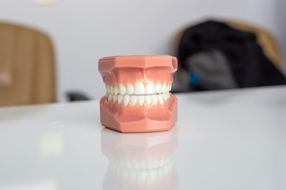
Numbering Systems in dentistry serve as abbreviations, instead of writing out the entire name of a tooth, such as permanent maxillary right central incisors.
It is as simple to assign it a number, letter, or symbol, such as #8 for the universal numbering system.
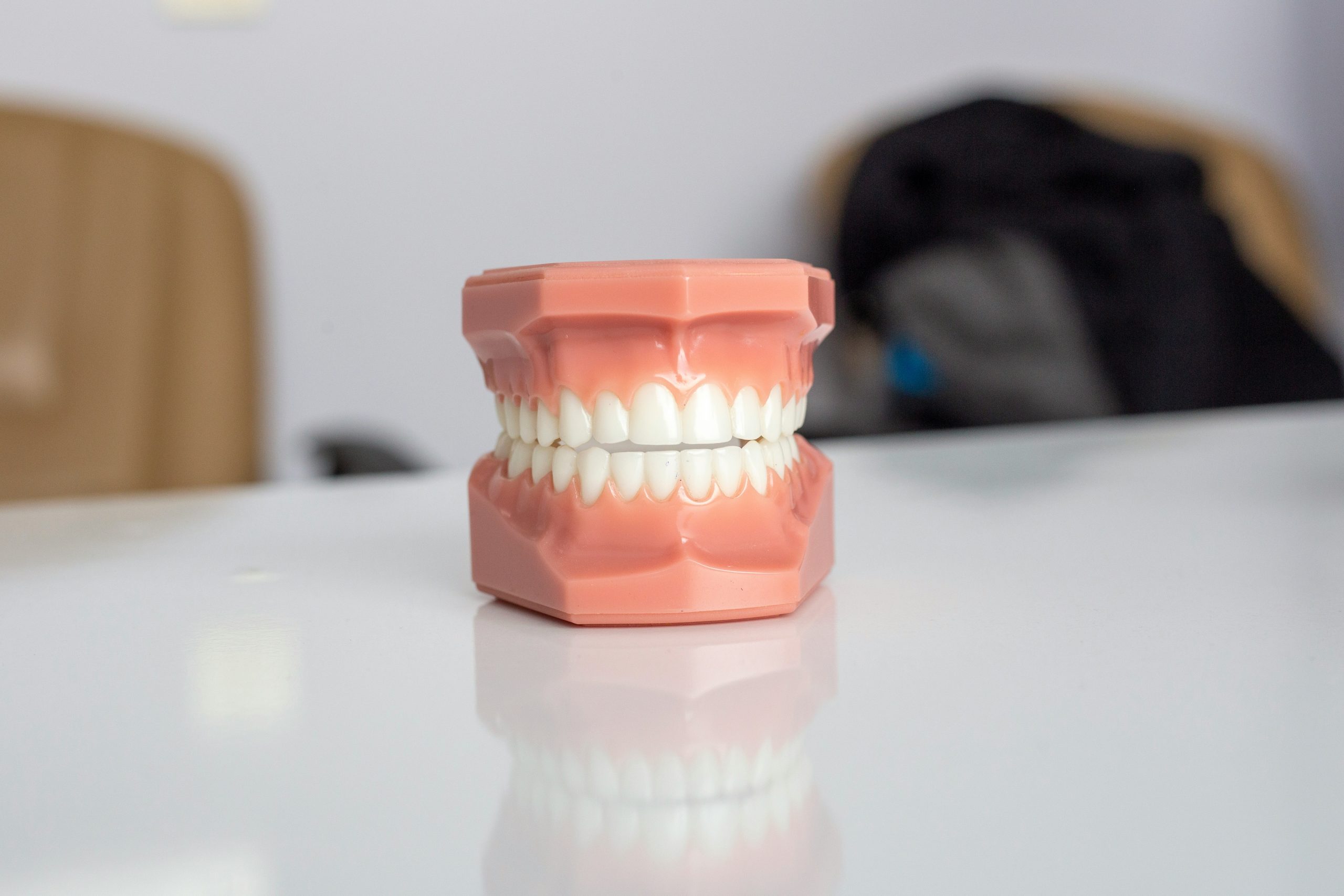
It employs a different number (1-32) in a consecutive arrangement for all permanent teeth and a number-letter (1 day to 20 days) for each of the deciduous teeth. You can read more similar posts on our medicine and health page of the site.
Permanent Teeth
The Universal numbering system assigns a specific number to each permanent tooth. The upper right third molar is #1, the upper right second molar is #2, and so forth around the entire maxillary arch to the upper left third molar, which is #16.
Since there are no more permanent teeth in the maxillary arch, the succession drops to the lower left third molar, which is #17, and continues around the entire mandibular arc, where the lower right third molar is #32. For example, tooth #11 is the permanent maxillary left canine.
Deciduous Teeth
The twenty teeth of the deciduous dentition are numbered in the same manner as the permanent teeth(1-20), except that a small (d) is added as a suffix to each number to designate deciduous.
The deciduous upper right second molar is thus #1d, while the upper left second molar is#10d. The lower right cani, for example, is #18d.
The most common system in use today for the design of deciduous teeth uses the capital letters(A through T).
The maxillary right deciduous second molar is tooth A, and the order progresses in the manner used with the 1-32 system for permanent teeth, so that the mandibular right deciduous second molar is tooth T.
Palmer notation method
Another commonly used numerical and letter notation scheme for identifying an individual tooth utilises a simple symbol, which differs for each of the four quadrants.
In addition, the numbers 1 through 8 are used to identify permanent central incisors through the third molar in the specified quadrant. Letters A through E with the quadrant symbol are used for the deciduous dentition.
There are Maxillary and Mandibular on the Right and Left.
Specific examples are:
6-Permanent maxillary right first molar
3-Permanent maxillary left canine
B-Deciduous mandibular left lateral incisor
4-Permanent mandibular right first premolar
FDI system
It is a simple binomial system, which includes both permanent and deciduous teeth. The first of the two numbers identifies the quadrant and whether the tooth is permanent or deciduous as follows:
1-Permanent maxillary right quadrant
2-Permanent maxillary left quadrant
3-Permanent mandibular left quadrant
4-Permanent mandibular right quadrant
5-Deciduous maxillary right quadrant
6-Deciduous maxillary left quadrant
7-Deciduous mandibular left quadrant
8-Deciduous mandibular right quadrant
The second number identifies the particular tooth in the quadrant, exactly like the Palmer notation method for permanent teeth(1-8).
The Deciduous
The deciduous teeth in each quadrant are numbered(1-5), the number increasing in size from the midline posteriorly.
The following are examples in notation the FDI system notation:
18-Permanent maxillary right third molar
27-Permanent maxillary left second molar
36-Permanent mandibular left first molar
45-Permanent mandibular right second premolar
54-Deciduous maxillary left canine
63-Deciduous maxillary left canine
72-Deciduous mandibular left lateral incisors
81-Deciduous mandibular right central incisors
The international system for permanent teeth
First Digit = quadrant
1/42/3
Example (18) can be analysed as follows;
1-The first indicates that the tooth is located in the permanent maxillary right quadrant.
8-The second number indicates that the tooth is eight from the midline and thus is a molar
Tooth formation
All deciduous teeth arise from the dental lamina. Later, the permanent successors arise from its lingual and distal extensions
Consider the following terms:
pdy, cap, enamel, bud, stage, dentin, epithelium, dental, stage, enamel knot, placode, chyme, dental-, ameloblasts, root, mesen-, dental mesenchyme, papilla, pulp, bone, odontoblasts, dental lamina, bud, cap, bell, eruption, initiation, morphogenesis, cell differentiation and matrix secretion.
Dental Lamina
At about the 7th week, the primary epithelial band is divided into an inner (lingual) process called the dental lamina and an outer (buccal) process called the Vestibular lamina.
ENAMEL ORGAN, also known as the dental organ, is a cellular aggregation seen in histologic sections of a developing tooth. Total activity of the ental lamina exceeds at least 5 years. As the teeth continue to develop, they lose their connection with the Dental lamina.
Clinical Considerations
XANODONTIA-Anodontia, also called anodontia vera, is a rare genetic disorder characterised by the congenital absence of all primary or permanent teeth.
Itis of 2 types(a)Complete anodontia/total anodontia (b) Partial anodontia/Sub-total anodontia
When classified by position, a supernumerary tooth may be referred to as
- A mesiodens
- A paramour
- A dissimilar
Vestibular Lamina
Where there is the Labial and buccal to the dental lamina in each dental arch, another epithelial thickening will develop solely because it is the vestibular lamina. This is termed a lip furrow band, which forms the oral vestibule between the jaws, the lips, and the cheeks.
Tooth Development
Each of these growths from the dental lamina represents the beginning of the enamel organ of the tooth bud.d
Dental Papilla
The tissue appears denser than the surrounding mesenchyme and represents the beginning of the dental papilla.
Developmental Stages
MORPHOLOGICAL
- Dental lamina
- Bud stage
- Cap stage
- Early bell stage
- Advanced bell stage
- Formation of enamel and dentin matrix
Physiological
- Initiation
- Proliferation
- Proliferation
- Histodifferentiation
- Morphodifferentiation
- Opposition
Bud Stage
The initial stage of tooth formation. The ectomesenchymal condensation that surrounds the tooth bud and the dental papilla is the tooth sac. The dental papilla, as well as the dental ssacare not well defined in the bud stage.
The cells of the dental papilla form the dentin, and pulpule the dental sac forms cementum and periodontal ligament.
Outer And Inner Enamel Epithelium
The cells in the concavity of the cap become tall columnar cells and represent epithelium. The outer enamel epithelium is separated from the dental sac and the inner enamel epithelium from the dental papilla by a delicate basement membrane.r ane
Stellate Reticulum
As the star-shaped cells form a cellular network, they are called the stellate reticulum. Study the terms below.
Ectoderm
Epithelium
Basal layer
Basement
membrane
Tooth germ
Stellate reticulum
Dental organ
Mesoderm
Dental sac
Dental papilla
Mesenchymal
The Enamel
The enamel knot acts as a signalling centre, as many important growth factors are expressed by the cells of the enamel knot and thus play an important role in determining the shape of the tooth. The dental papilla and the dental sac are pronounced during this stage of dental development. lopment
Bell Stage
Due to the continued growth of the enamel, el organelle acquires a bell shape. In the bell stage, the crown shape is determined. The folding of enamel organs to cause different crown shapes is shown to be due to different rates of mitosis and differences in cell differentiation. Timeme
Dental Sac
The dental sac shows a circular arrangement of fibres and resembles a capsule around the en. The organ, the fibre of the dental sac, forms the periodontal ligament fibres that span between the root and the bone. The junction between the inner enamel epithelium and odontoblasts outlines the future dentino-enchymal junction.n
Advanced Bell Stage
- Characterised by the commencement of mineralisation and root formation
- Formation of dentin occurs first as a layer along the future dentino-enamel junction in the region of future cusps
- After the first layer of dentin is formed, the ameloblasts lay down enamel over the dentin in the future incisal and cuspal areas
- The enamel formation then proceeds coronally and cervically in all the regions from the dentin-enamel junction towards the surface.
- The cervical position of the enamel organ gives rise to her HERS epithelial root sheath of the hair.
